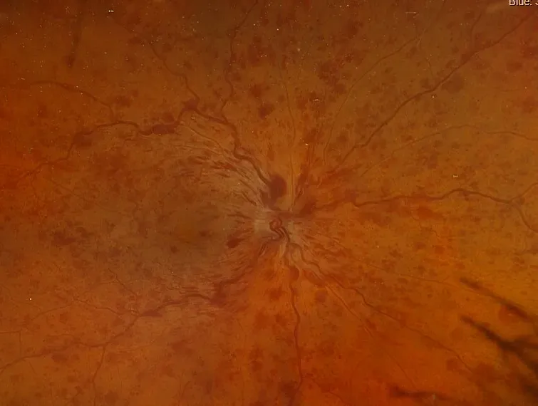
A retinal vein occlusion can interfere with the normal flow of blood out of your retina, leading to bleeding, swelling, and changes in vision. At Greater Dallas Retina in McKinney, Texas, Kruti Dajee, MD, Archana Nair, MD, and the team diagnose and treat retinal vein occlusions using detailed imaging and treatments like intravitreal injections. Contact the Greater Dallas Retina team if you experience sudden or unexplained changes in your sight to arrange a retinal vein occlusion evaluation. Call the office or schedule a consultation online today.
Retinal Vein Occlusion Q & A
What happens during a retinal vein occlusion?
Retinal veins carry blood away from the light-sensitive retina at the back of your eye. A retinal vein occlusion (RVO) develops when one of these veins becomes blocked, preventing blood from draining normally.
Pressure then builds within smaller retinal blood vessels. They can leak blood and fluid into surrounding tissue, potentially affecting vision.
There are two main types of retinal vein occlusion. Branch retinal vein occlusion (BRVO) is the more common type, affecting one of the smaller veins draining part of the retina. Central retinal vein occlusion (CRVO) affects the main retinal vein and can involve a much larger area.
Why do retinal veins become blocked?
Retinal vein occlusions often develop where a hardened or thickened retinal artery presses against a nearby vein. This can interfere with blood flow and contribute to clot formation.
Risk increases with age and is associated with vascular conditions such as:
- High blood pressure
- Diabetes
- High cholesterol
- Cardiovascular disease
Glaucoma can also increase the risk of some retinal vein occlusions. For that reason, identifying an RVO may prompt an evaluation of your wider cardiovascular risk as well as treatment of your eye.
How does imaging assess retinal vein occlusion?
The Greater Dallas Retina team examines your retina through a dilated pupil to look for characteristic bleeding, swollen vessels, and other changes.
Optical coherence tomography (OCT) provides detailed cross-sectional images of the retina and can show whether fluid has accumulated within the macula.
Fluorescein angiography can provide further information about retinal circulation. It helps identify areas where blood flow has become restricted, locate leaking vessels, and assess whether abnormal new blood vessels are developing.
What treatments help with retinal vein occlusion?
It’s generally not possible to remove the original blockage. Instead, treatment concentrates on controlling its effects and protecting the retina from further damage.
You might need intravitreal anti-VEGF injections if RVO causes macular edema. These medications reduce vascular leakage and swelling and may need to be repeated over time. Steroid treatment is another option in selected cases.
Some people develop abnormal new blood vessels because areas of the retina aren’t receiving enough oxygen. These vessels can bleed and may contribute to complications, including vitreous hemorrhage or increased eye pressure. Laser treatment may be necessary when abnormal vessel growth develops.
If you experience sudden painless blurring, distortion, or loss of vision, call Greater Dallas Retina or request an appointment online today.